Video imaging used to identify effectiveness of rituximab on cancer
Video microscopy has been used to identify why a cancer treatment is so effective.
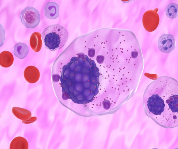
High quality video imaging has been used to uncover why the drug rituximab is so effective at killing B cell malignancies, such as lymphoma and leukaemia.
The study, from the Manchester Collaborative Centre for Inflammation Research in the UK, explained how video footage enabled the team to see how the drug worked.
Rituximab tended to stick to one side of the cancer cell, drawing a cluster of protein molecules to that side.
When the natural killer cells latched onto the rituximab cap in the B cell, it had an 80 per cent success rate at killing the cell, compared to 40 per cent when the B cell did not have a cluster of protein molecules on one side.
"It was only possible for us to unravel the mystery of why this drug was so effective, though the use of video microscopy," commented Professor Daniel Davis.
He added that this ability to polarise a cell by moving proteins within it should be considered when new antibodies are being tested as potential treatments for cancer cells.
The results have been published online by the journal Blood.
Related News
-

News BioNTech to begin mRNA vaccine manufacturing in Rwanda by 2025
German biotechnology company BioNTech has stated their intentions to begin production at their mRNA vaccine factory in Rwanda by 2025, which will mark the first foreign mRNA vaccine manufacturing site on the continent of Africa. -

News Identifying Alzheimer’s Disease biomarker proteins with whole blood tests
A University of Manchester spin-out pharmaceutical company, PharmaKure, has reported successful study results for the quantification of Alzheimer’s Disease biomarker proteins with a whole blood test. -

News Bill & Melinda Gates Foundation to boost mRNA vaccine initiatives in Africa with USD $40m
To address vaccine inequality and accessibility issues, the Bill & Melinda Gates Foundation aims to deliver USD $40m to various biotech companies and vaccine manufacturers in support of mRNA vaccine development. -

News CPHI Podcast Series: Exploring neurological frontiers in Alzheimer's and beyond
The next episode of the CPHI Podcast Series delves into the science and background behind some recent developments in the field of Alzheimer's disease and neurological disorders. -

News Is patient centricity the future of pharmaceutical manufacturing?
In this interview with Sandra Sánchez y Oldenhage, President of PharmAdvice, she speaks to the importance of considering patients in the manufacturing stages of the pharmaceutical supply chain, and how it can redefine healthcare. -

News CPHI Podcast Series: How to leverage AI for Drug Discovery
Artificial intelligence is the topic of debate in the latest episode from the CPHI Podcast Series, where Digital Editor Lucy Chard speaks with Bill Whitford of DPS Group about the integration of AI in healthcare. -
News Pfizer forges ahead with blood cancer therapy after approval from FDA
Pfizer gains accelerated approval from the US FDA for their new bispecific antibody therapy for multiple myeloma, set to address an unmet need for patients. -

News Alzheimer's drug donanemab deemed effective in landmark clinical trial
Results from the TRAILBLAZER-ALZ 2 Randomised Clinical Trial into the use of donanemab to treat early symptoms of Alzheimer’s disease have been analysed.
Position your company at the heart of the global Pharma industry with a CPHI Online membership
-
Your products and solutions visible to thousands of visitors within the largest Pharma marketplace
-
Generate high-quality, engaged leads for your business, all year round
-
Promote your business as the industry’s thought-leader by hosting your reports, brochures and videos within your profile
-
Your company’s profile boosted at all participating CPHI events
-
An easy-to-use platform with a detailed dashboard showing your leads and performance