The Curtain Rises On DNA–Protein Interactions
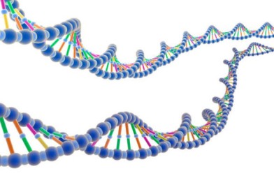
For the first time, scientists have watched proteins interact with long single-stranded DNA molecules in real time.The new method should yield insights into DNA replication and repair, the researchers say.
For the first time, scientists have watched proteins interact with long single-stranded DNA molecules in real time (Anal. Chem., DOI: 10.1021/ac302117z). The new method should yield insights into DNA replication and repair, the researchers say.
Until now, scientists could only watch proteins interacting with double-stranded DNA or very short single strands, says Eric Greene of Columbia University, who developed the new method. But, he says, “Basically anything to do with DNA metabolism or DNA repair involves single-stranded DNA intermediates.”
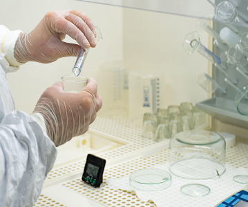
To find a way to monitor long single strands, Greene built upon a method his team devised six years ago, which visualizes proteins binding to double-stranded DNA (Langmuir, DOI: 10.1021/la051944a). They tethered fluorescent DNA molecules to lipids in a bilayer that coated the surface of a microfluidic sample chamber. When the researchers flowed buffer through the chamber, the DNA molecules diffused within the bilayer, eventually aligning along a nanofabricated barrier in the bilayer. The researchers added proteins to the chamber and observed their interactions with DNA in real time by noting changes in the DNA’s fluorescent signal. The team called the method “DNA curtains” because the hundreds of aligned DNA strands—visualized by total internal reflection fluorescence microscopy—reminded them of a window curtain.
But the technique didn’t work with single-stranded DNA, which is so flexible that it often folds upon itself into extensive secondary structures that preclude protein binding. Also, the intercalating fluorescent dyes that label double-stranded DNA can break single strands of DNA.
So Greene and his coworkers found a way to simultaneously unravel folded-up single-stranded DNA and fluorescently label it. The key was a protein called replication protein A (RPA), which binds single strands during DNA replication to prevent them from folding up. When the researchers added a fluorescently labeled version of the protein to a DNA-curtains assay, it coated single-stranded DNA molecules, helping extend the DNA and keep it accessible to proteins. Because RPA coats all single-stranded DNA in cells, the researchers say, the ubiquitous protein is unlikely to interfere with the binding of other proteins.
To test whether the assay could visualize interactions of single-stranded DNA with other proteins, the team injected fluorescently tagged Sgs1 protein into a flow cell containing RPA-bound DNA curtains. Sgs1 is a yeast helicase protein involved in DNA repair. Using fluorescence microscopy to visualize the labeled RPA and Sgs1 proteins, the researchers watched Sgs1 bind single-stranded DNA in real time.
Greene says that the new assay will allow researchers to probe when and where proteins bind single-stranded DNA, and how multiple proteins influence each other’s binding. “We can watch all of this stuff as it happens,” he says.
Maria Spies of the University of Iowa says, “The DNA curtains approach combines the benefits of single-molecule and bulk studies.” That’s because, she explains, researchers can use it to visualize hundreds of single-stranded DNA molecules at the same time. “I can’t wait to try this system in my lab,” she says.
Related News
-

News CPHI Frankfurt 2022: Innovator Interview – DSM Biomedical
At CPHI Frankfurt we spoke to Anne-Cecile Bayne, Global Science & Innovation Lead Pharma and Medical Nutrition, and Marc Hendriks, Vice President Strategy & Business Development, on their expertise in nitrosamines and business strategy at DSM Biomedica... -

News New WHO health emergency guidelines expect full transparency from Big Pharma
The WHO are proposing a new set of pandemic guidelines to set out how future global health crises should be handled. -
.png)
News Magic mushrooms could be used to treat mental health conditions
A compound found in magic mushrooms, psilocybin, could be used to treat mental health conditions and help patients suffering with severe depression, as shown by the results of the largest study of its kind to date. -

News UK-based partnership to launch DETERMINE study into rare cancer research
UK-based CRO Quanticate is set to partner with Cancer Research UK for the launch of the DETERMINE study focused on testing a range of existing and approved drugs and therapies on rare cancers. -

News FDA approves Thermo Fisher blood tests for wheat and sesame allergies
Both tests have been approved by the US regulator for in vitro diagnostic use -
News QIAGEN launches world’s first syndromic test for monkeypox
The test can distinguish between monkeypox and other diseases that cause similar symptoms. -

News Monkeypox Update: Vaccine shortage, sewage surveillance and global testing
As concern over the monkeypox outbreak continues to rise, we take a look at major developments from the first week of August. -

News CPHI Podcast Series: The importance of novel excipients for innovative drug development
The latest episode in the CPHI Podcast Series dives into the world of novel excipients and explores their importance for innovative drug development.
Position your company at the heart of the global Pharma industry with a CPHI Online membership
-
Your products and solutions visible to thousands of visitors within the largest Pharma marketplace
-
Generate high-quality, engaged leads for your business, all year round
-
Promote your business as the industry’s thought-leader by hosting your reports, brochures and videos within your profile
-
Your company’s profile boosted at all participating CPHI events
-
An easy-to-use platform with a detailed dashboard showing your leads and performance